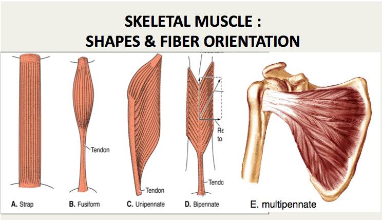
muscle fiber orientation
Measurements using common surface. Knowledge of skeletal muscle fiber orientations is important for modeling mechanical properties and behavior of muscle tissue.

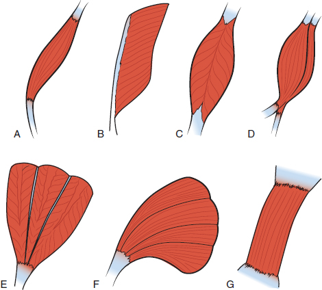
Pennate Muscle An Overview Sciencedirect Topics
FO fibers use aerobic metabolism to produce ATP but produce higher tension.
. The metabolite profile changes due to the muscle fiber orientation demonstrate that the positioning potentially causes inaccuracy in 1 H-MRS spectrum analysis. The dense muscle fiber microstructure gives rise to orientation dependent mr features with anisotropic overall motion of the creatine cr and phosphocreatine pcr molecules causing residual dipolar couplings first described for the total observed creatine tcr crpcr resonances 1 2 while orientation dependence was later also reported for. Illustrations of muscle fiber orientation were done and the assumed location of motor endplate bands marked.
Tending to approach each other. The angle of inclination of muscle fibers from coronal section was largest in the innermost and outermost zones and was progressively diminished toward the middle layer in all the hearts. An oxygen debt is created as a result of muscle use.
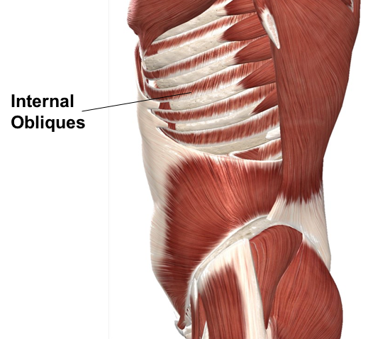
Results showed that the fibres of obliquus externus abdominis were about 4 degrees more vertical than the lower edge of the. Thirty-seven embalmed cadavers 19 males and 18 females were examined. Muscle fiber orientation MFO is an important parameter related to musculoskeletal functions.
Muscle fiber orientation curvature and length is known to change with changes in force within the muscle Herbert et al. This study aimed to measure muscle fibre orientation and other parameters of muscle morphology of the abdominal muscles in relation to palpable bony landmarks. Home Browse by Title Proceedings 2015 IEEE International Conference on Image Processing ICIP An automatic muscle fiber orientation tracking algorithm using Bayesian Kalman Filter for ultrasound images.
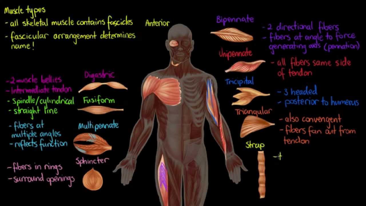
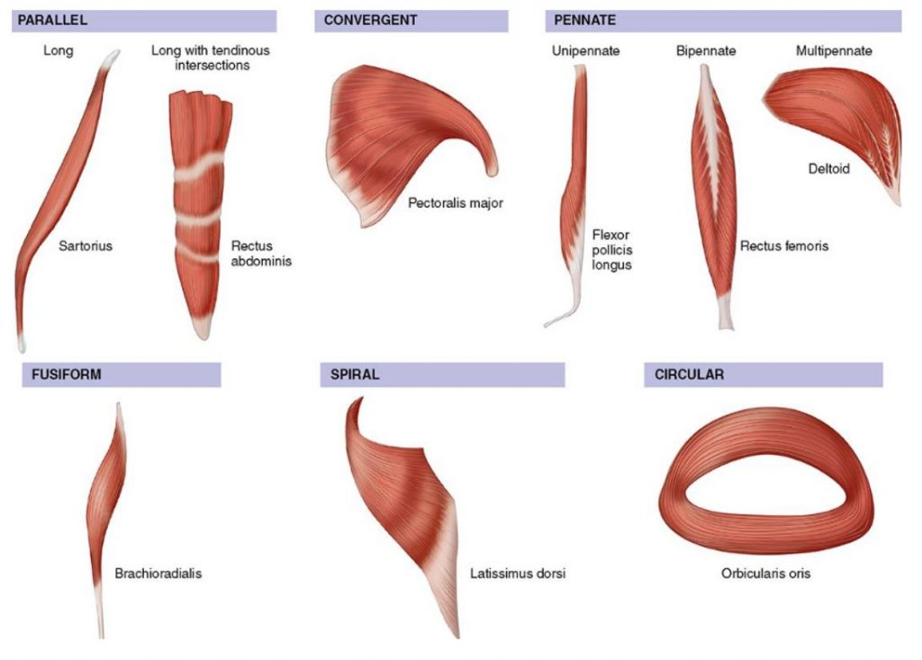
Muscle fiber orientation MFO is an important parameter related to musculoskeletal functions. Examples include Sartorius and Sternocleidomastoid. The muscle fibers in native skeletal muscle are closely packed together in an extracellular matrix ECM to form an organized tissue with high cell density and three-dimensional 3D cellular orientation.
Analysis the orientation of muscle fibers in planarians. The automatic methods proposed in recent years also involved voting procedures which were computationally expensive. The automatic methods proposed in recent years also involved voting procedures which were computationally expensive.
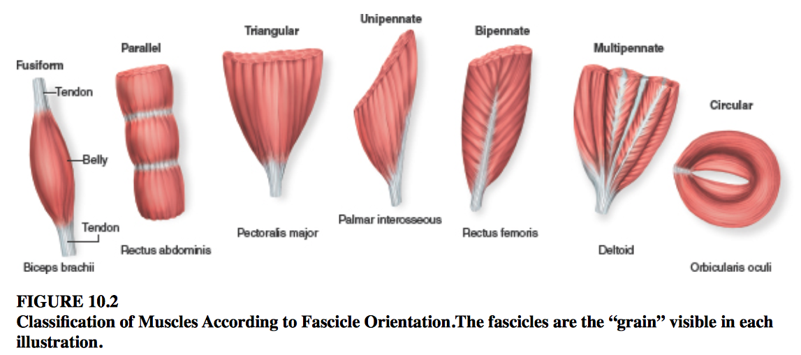
Some textbooks include Fusiform muscles in the parallel group. The proximal and distal musculotendinous junctions in muscles of the upper and lower extremities were identified. This study aimed to measure muscle fibre orientation and other parameters of muscle morphology of the abdominal muscles in relation to palpable bony landmarks.
The traditional manual method for MFO estimation in sonograms was labor-intensive. This study was designed to locate the middle of the muscle fibers of commonly injected muscles thus identifying the endplate zone of these muscles. Thirty-seven embalmed cadavers 19 males and 18 females were examined.
The three types of muscle fiber are slow oxidative SO fast oxidative FO and fast glycolytic FG. Results showed that the fibres of obliquus externus abdominis were about 4more vertical than the lower edge of the eighth rib. In the inner layer the inclination was.
An automatic muscle fiber orientation tracking algorithm using Bayesian Kalman Filter for ultrasound images. Analysis the orientation of muscle fibers in planarians - GitHub - walkernoreenmuscle_fiber_orientation. The traditional manual method for MFO estimation in sonograms was labor-intensive.
SO fibers use aerobic metabolism to produce low power contractions over long periods and are slow to fatigue. A skeletal muscle fiber arrangement in which the fibers are in an somewhat parallel orientation spread across a wide bone surface at the origin and coming together at a tendon attachment at the insertion. Two groups of muscle fibers which are located within the deep perineal pouch and surround the urethra are called the urethral sphincter Oelrich 1980 The urethral sphincters are used to control the exit of urine in the urinary bladder through the urethra mainly the external urethral sphincter which is under voluntary control.
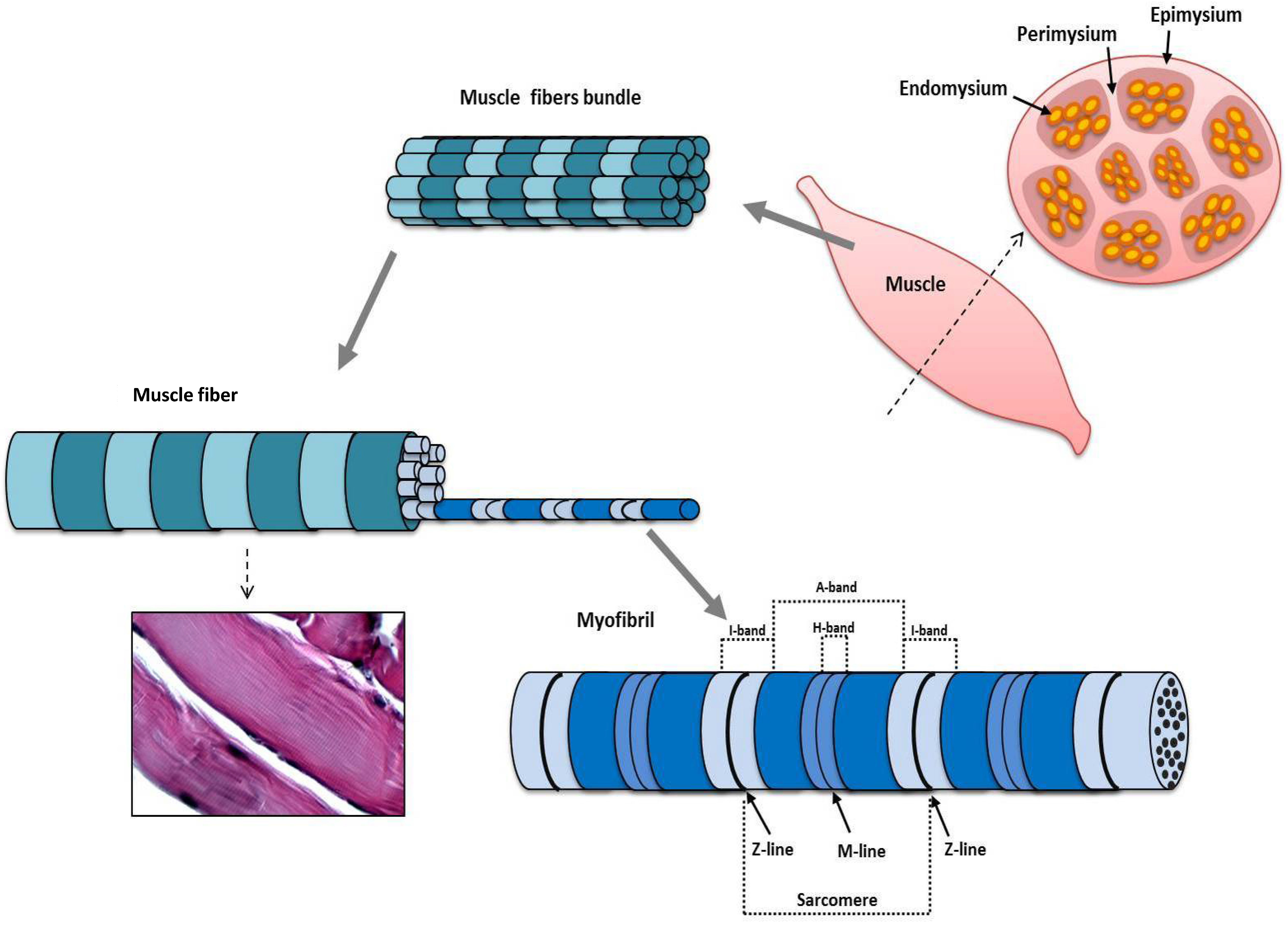
Muscle fiber orientation STUDY PLAY parallel strap have fibres which as the name suggests run parallel to each other. The present findings indicates that 1 the right ventricular hypertrophy induced by pressure loading is characterized not only by an increase in ventricular weight and muscle fiber thickness but also by a change i n intramyocardial fiber orientation and 2 the hypertrophic right ventricle can regress both functionally and morphologically to a normal state after removal of the. These tissues include the skeletal muscle fibers blood vessels nerve fibers and connective tissue.
This study reveals that the muscle orientation at 0 30 60 and 90 to the main magnetic field significantly affects the metabolite profile and quantification. The endplate zone is assumed to be at about the midpoint of a muscle fiber. The ECM plays an essential role in the growth attachment alignment and differentiation of myoblasts and is part of the signaling mechanism involved in myogenesis.
The proximal and distal musculotendinous junctions in muscles of the upper and lower extremities were identified. This study was designed to locate the middle of the muscle fibers of commonly injected muscles thus identifying the endplate zone of these muscles. Each skeletal muscle is an organ that consists of various integrated tissues.
They are normally long muscles which cause large movements are not very strong but have good endurance. Orientation of muscle fibers was determined. This fiber orientation provides a good compromise in providing for both range of motion and power for.
Comparisons between CFD and Diffusion Tensor Imaging. CFD Simulations for 3D Muscle Fiber Architecture in Finite Element Analysis. Muscle fiber orientation in the left ventricular myocardial layer was histometrically estimated in normal concentric and eccentric hypertrophied hearts.
Color illustrations will be shown. Tending to one point of focus. Broadly there are two main approaches to extracting fiber orientationcurvature automatically from ultrasound.
With the thought that the endplate zone is at the middle of the muscle fiber this detailed study of muscle fibers helps identify assumed location of motor endplates of specific muscles thereby improving technique. Each skeletal muscle has three layers of connective tissue that enclose it provide structure to the muscle and compartmentalize the muscle fibers within the muscle.
Imaging Muscle Radiology Key
How Muscles Work Part 2 Of 2 Shapelog

Classification Of Muscles Based On The Directions Of Muscle Fibers The Download Scientific Diagram
Jfmk Free Full Text Morphological And Functional Aspects Of Human Skeletal Muscle Html
Muscle Histology Flashcards Chegg Com
Muscle Types Youtube
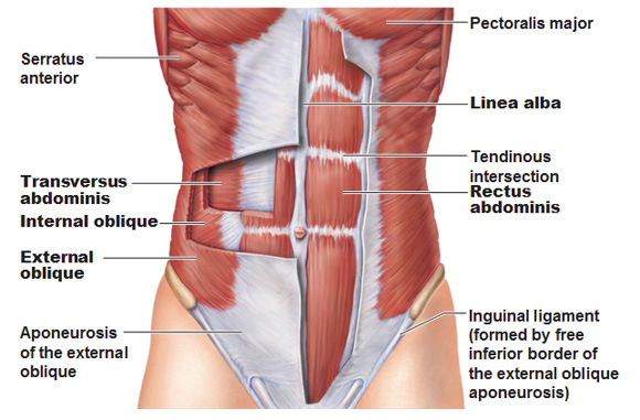
Muscles Of The Abdominal Wall

Organization Of Skeletal Muscles Course Hero

Pennate Muscle An Overview Sciencedirect Topics
Changing The Way You Learn Flashcards

Myocardial Fiber Orientation And Direction Of Rotation Myocardial Download Scientific Diagram

Organization Of Skeletal Muscles Course Hero

Skeletal Muscle Shapes Fusiform Muscles Thick In Middle And Tapered At Ends Parallel Muscles Have Parallel Muscle Fibers Convergent Muscle Broad At Ppt Download

3 Orientation Of Cardiac Muscle Fibers Download Scientific Diagram
2
Muscles Advanced Anatomy 2nd Ed

Myocardial Mechanics Structure And Function Of Myocardial Fibers Ecg Echo

11 2 Explain The Organization Of Muscle Fascicles And Their Role In Generating Force Anatomy Physiology

Fascicle An Overview Sciencedirect Topics
0 Response to "muscle fiber orientation"
Post a Comment